


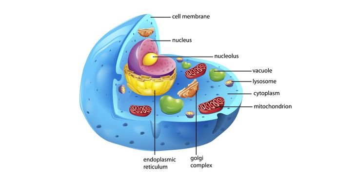
A labeled animal cell provides one of the most essential visual tools for students, educators, and biology enthusiasts seeking to grasp the complex inner workings of eukaryotic cells. Unlike plant cells, animal cells lack a rigid cell wall and chloroplasts, giving them a more flexible structure that supports diverse functions from movement to specialized tissue formation. Exploring a labeled animal cell reveals an intricate world of organelles working in harmony to sustain life, making it a foundational concept in biology education and research.
This guide offers a thorough examination of the labeled animal cell, breaking down each component’s role, significance, and interactions within the cellular environment. Whether you are preparing for an exam, teaching a class, or simply satisfying your curiosity about how life operates at the microscopic level, understanding the labeled animal cell opens doors to appreciating the remarkable complexity packed into every living creature.
The Overall Structure of a Labeled Animal Cell
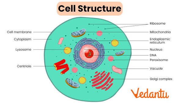
When examining a labeled animal cell, the first impression is often the cell membrane, a thin, flexible boundary that separates the cell’s internal environment from the outside world. This phospholipid bilayer not only protects the cell but also regulates what enters and exits, maintaining the delicate balance necessary for survival. In diagrams of a labeled animal cell, the membrane appears as a distinct outer line, sometimes with small proteins embedded within it that facilitate communication and transport.
Moving inward, the cytoplasm fills the space between the membrane and the nucleus. This gel-like substance serves as the cell’s internal environment where many chemical reactions occur. In a well-drawn labeled animal cell, you will notice various organelles suspended within this cytoplasmic matrix, each performing specialized tasks that contribute to the cell’s overall function. The dynamic nature of the cytoplasm allows organelles to move and interact efficiently, supporting the constant activity required for life processes.
The most prominent feature in any labeled animal cell is typically the nucleus, often positioned near the center. This control center houses the cell’s genetic material in the form of chromatin, which condenses into chromosomes during cell division. The nuclear envelope, complete with nuclear pores, allows selective exchange of materials between the nucleus and cytoplasm. Understanding the nucleus in the context of a labeled animal cell helps explain how genetic information directs cellular activities and ensures proper replication.
Key Organelles in a Labeled Animal Cell
Mitochondria stand out in any labeled animal cell diagram as bean-shaped structures with inner folds called cristae. Often referred to as the powerhouses of the cell, these organelles generate adenosine triphosphate through cellular respiration, providing the energy necessary for all cellular functions. Their abundance in high-energy cells like muscle fibers highlights their critical importance. When studying a labeled animal cell, recognizing the mitochondria helps explain why certain tissues require more energy and how disruptions in mitochondrial function can lead to various health conditions.
The endoplasmic reticulum appears as a network of interconnected membranes in a labeled animal cell. The rough endoplasmic reticulum, studded with ribosomes, specializes in protein synthesis and modification, while the smooth variety focuses on lipid production and detoxification. This distinction becomes clear in detailed diagrams of a labeled animal cell, where the presence or absence of ribosomes creates visible differences in texture. The endoplasmic reticulum works closely with other organelles to ensure proteins and lipids reach their proper destinations, making it a crucial component of the cellular logistics system.
Another vital structure visible in a labeled animal cell is the Golgi apparatus, which modifies, sorts, and packages proteins and lipids for storage or transport. Its stacked, flattened sacs give it a distinctive appearance that students quickly learn to identify. The Golgi apparatus acts as a cellular post office, ensuring molecules reach their correct locations either within the cell or for secretion outside. This organelle’s role becomes particularly important when considering how cells communicate with their environment and maintain internal organization.
Specialized Structures and Their Functions
Centrioles often appear near the nucleus in a labeled animal cell, typically arranged in pairs within the centrosome. These cylindrical structures play a crucial role in organizing microtubules during cell division, helping ensure accurate chromosome separation. While not all animal cells display prominent centrioles in every stage, their presence in a labeled animal cell diagram reminds us of the precise mechanisms governing cellular reproduction.
Lysosomes, the cell’s digestive compartments, contain enzymes that break down waste materials and cellular debris. In a labeled animal cell, they appear as small, membrane-bound vesicles. Their function proves essential for maintaining cellular cleanliness and recycling components, preventing the accumulation of harmful substances that could damage the cell over time.
The cytoskeleton, though sometimes less emphasized in basic labeled animal cell diagrams, provides structural support and enables movement. Composed of microfilaments, intermediate filaments, and microtubules, this internal framework maintains cell shape while facilitating transport of materials within the cytoplasm. Understanding the cytoskeleton in relation to a labeled animal cell helps explain how cells achieve complex movements and maintain integrity under mechanical stress.
Comparing Animal Cells with Other Cell Types
When studying a labeled animal cell, contrasting it with plant cells reveals fascinating differences that reflect their distinct lifestyles. Plant cells possess rigid cell walls made of cellulose, chloroplasts for photosynthesis, and large central vacuoles for storage and turgor pressure. Animal cells, lacking these features, rely on their flexible membranes and diverse organelles to adapt to various environments and functions within multicellular organisms.
This comparison becomes particularly educational when examining a labeled animal cell alongside its plant counterpart. The absence of a cell wall gives animal cells greater flexibility, allowing for specialized shapes and movements essential for functions like muscle contraction or immune responses. Meanwhile, the presence of centrioles in animal cells supports more dynamic cell division processes compared to plant cells, which use different mechanisms for organizing their mitotic spindles.
Educational Value and Learning Applications
A well-designed labeled animal cell serves as an invaluable educational tool across different learning levels. For beginners, it provides a clear visual reference for memorizing organelle names and basic functions. More advanced students use it to understand interactions between cellular components and how disruptions in specific structures can lead to diseases.
Teachers often incorporate labeled animal cell diagrams into lessons through various methods, from simple identification exercises to complex analysis of cellular processes. Interactive digital versions allow students to explore different layers and functions, making abstract concepts more concrete and memorable. The visual nature of these diagrams helps accommodate different learning styles, ensuring broader comprehension among diverse student populations.
Practical Applications in Research and Medicine
Beyond education, understanding the labeled animal cell has significant implications for scientific research and medical applications. Researchers studying cellular mechanisms rely on accurate models of animal cell structure to design experiments and interpret results. In medical fields, knowledge of cellular anatomy helps explain disease processes and develop targeted treatments.
For example, understanding mitochondrial function in a labeled animal cell has led to advances in treating metabolic disorders. Similarly, research into the endoplasmic reticulum’s role in protein folding has contributed to understanding diseases like Alzheimer’s and certain cancers. These practical applications demonstrate how fundamental knowledge of cellular structure translates into real-world benefits for human health and scientific progress.
Conclusion
The labeled animal cell represents far more than a simple biology diagram. It serves as a window into the remarkable complexity and coordination that sustains all animal life. From the protective cell membrane to the energy-producing mitochondria and the information-storing nucleus, each component works in harmony to maintain the delicate balance necessary for survival. As we continue advancing our understanding of cellular biology, the labeled animal cell remains a fundamental teaching tool that bridges basic education with cutting-edge research. Whether you are a student beginning your scientific journey, an educator sharing knowledge with others, or simply a curious individual seeking to understand life at its most basic level, exploring a labeled animal cell offers profound appreciation for the intricate systems that make us who we are. The more we study and understand these cellular foundations, the better equipped we become to address health challenges, develop new technologies, and marvel at the elegance of biological organization.
FAQ
What is a labeled animal cell? A labeled animal cell is a diagram that identifies and names the key organelles and structures within an animal cell, helping students and researchers understand cellular anatomy.
What is a labeled animal cell?
A labeled animal cell is a diagram that identifies and names the key organelles and structures within an animal cell, helping students and researchers understand cellular anatomy.
What are the main parts of a labeled animal cell?
The primary components include the cell membrane, nucleus, mitochondria, endoplasmic reticulum, Golgi apparatus, ribosomes, lysosomes, and centrioles.
How does an animal cell differ from a plant cell?
Animal cells lack cell walls, chloroplasts, and large central vacuoles, but contain centrioles that aid in cell division.
Why is studying a labeled animal cell important?
It provides foundational knowledge for understanding biology, disease mechanisms, and how living organisms function at the cellular level.
Where can I find accurate labeled animal cell diagrams?
Reputable educational websites, biology textbooks, and scientific resources offer detailed and accurate diagrams for study purposes.
Read More:
Maplestar Dandadan: The Viral Fan Animator Bringing Dandadan’s Chaos to Life
